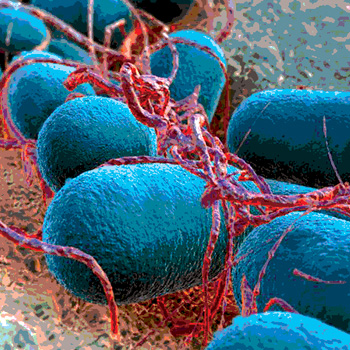
Shiga toxin-producing Escherichia coli (STEC) can cause illnesses that range from diarrhea to hemorrhagic colitis and hemolytic-uremic syndrome (HUS). More than 70 serotypes of STEC have been described, and fortunately, only a handful have been associated with these severe diseases. The STEC that can cause these severe diseases are referred to as Enterohemorrhagic E. coli (EHEC). E. coli O157:H7 is the EHEC most often associated with the severest forms of disease as well as outbreaks from contact with or consumption of contaminated foods, animals and water. It should be noted that the food has historically been ground beef; the animals, cattle; and the water presumably contaminated by runoff from a cattle production facility.
Numerous non-O157 EHEC have been linked to illnesses and outbreaks of disease similar to that of E. coli O157:H7, with only one occurrence linked to beef. The U.S. Centers for Disease Control and Prevention has identified the most common non-O157 EHEC and reported that just six serotypes (O26, O45, O103, O111, O121 and O145) are responsible for 71 percent of total infections from all sources. As part of its strategy to focus more on prevention, the U.S. Department of Agriculture Food Safety and Inspection Service (USDA FSIS) in the Federal Register implemented its decision to consider STEC of the six most frequent serogroup adulterants in certain beef products and began testing beef trimmings for these pathogens on June 4, 2012. In response, commercial test kit manufacturers and testing laboratories have rapidly introduced test kits and services that can detect and/or confirm the presence of the “Top Six” EHEC.
Detection of Top Six EHEC
In Chapter 5B of the FSIS Microbiological Laboratory Guidebook (MLG), the testing method used by FSIS to detect and confirm the presence of these adulterants is described. An initial polymerase chain reaction (PCR) screen targets the detection of stx and eae genes. If stx and eae are present, then the sample is considered reactive and taken forward to an additional round of PCR to determine if any of the Top Six O-groups are present. When stx, eae and an O-group are detected, the sample is considered a potential positive and goes forward to culture confirmation.
What are these things (stx, eae, and an O-group) and why are they the targets? The initial PCR screening targets the key virulence factors responsible for EHEC disease. To initiate an infection, EHEC must tightly bind to the intestinal lining, which is accomplished using a protein called intimin (expressed by the eae gene). Then the tightly bound EHEC produce Shiga toxins (expressed by the stx1 and/or stx2 genes) that damage an infected individual’s body. The second round of PCR targets genes that express enzymes used by nearly all E. coli to assemble various surface proteins. These surface proteins are antigenic and are used by laboratories to distinguish one O-group of E. coli from another.
Some E. coli may possess stx, whereas others may possess eae, and many others may be of one of the O-groups. Thus, it is not hard to imagine a situation where three different E. coli may be present that together provide all the targets for a potential positive test result. But would such a situation occur often enough to be of any consequence? Scientists at the U.S. Meat Animal Research Center in Clay Center, NE, asked that question of a set of nearly 4,000 ground beef samples in which they had cultured and identified only six non-O157 EHEC. They reported the answer in the October 2012 issue of Applied and Environmental Microbiology and state how they found that nearly 5 percent of the samples could be classified as potential positives and that only two of the six samples found to be contaminated by a Top Six EHEC were classified as a potential positive. Thus, the number and rate of potential positive samples can be significantly greater than the number of culture-confirmed EHEC, and the EHEC may be present and go undetected using this scheme.
Since there is no way when using stx, eae and O-groups as targets to know whether they are present in a single cell or multiple cells, alternative targets are under investigation. For instance, sequencing the O-group genes of numerous EHEC isolates was completed at the USDA Meat Animal Research Center that identified single nucleotide polymorphisms (SNPs) that correlate with the presence of stx and eae in an E. coli. Additional study of these organisms at the E. coli Reference Laboratory at the University of Santiago de Compostela in Lugo, Spain, has determined that EHEC of particular O-groups contain eae of specific subgroups. With minor exceptions, EHEC O26 contain eae-β, EHEC O145 and O157 contain eae-γ, EHEC O111 contain eae-θ and EHEC O45, O103 and O121 contain eae-ε. So if stx, eae-β and O45 are identified in a sample, it can be predicted that the eae-β (associated with EHEC O26) does not belong to the O45 organism detected, thus allowing what was a potential positive to be disqualified.
The correlation among eae subtypes extends beyond just that gene. When EHEC tightly bind to the intestinal wall using eae, they control and alter the cell they bind to by expressing and injecting effector molecules into the intestinal cell. These effector molecules are encoded by genes referred to as nle genes, because they are located in sections of the E. coli genome called pathogenicity islands rather than in the genes surrounding eae. A study by Brian Coombes illustrated the effect these molecules may play in predicting the pathogenesis of an EHEC. Coombes showed that as the number of these genes present in an isolate increased, so did the likelihood that the isolate was from either an outbreak or a case of HUS. From these studies, we can predict that nle genes may serve as targets for detection. At least one testing facility is using an nle gene in its protocol to identify the presence of a non-O157 EHEC.
Other EHEC-specific targets continue to be identified. One example is the EHEC hemolysin. A new test system under development aims to detect unique gene sequences associated with EHEC hemolysin to indicate the presence of EHEC. Another example is the recent work reported from the lab of Patrick Fach. His laboratory has identified two novel genes found in E. coli O157:H7 called Z2098 and Z2099 that are closely associated with EHEC and which they suspect may be useful markers for targeted detection of EHEC.
Current Detection Methods
With all the targets available to de-tect Top Six EHEC, there are many choices. Seven companies currently provide non-O157 EHEC testing or confirmation kits or services. One company provides an assay that has AOAC approval, while six others have received letters of no objection from FSIS as suitable replacements for the MLG assay until full AOAC approval is obtained. Three of these companies are marketing systems that follow the FSIS MLG methodology of identifying EHEC through the detection of stx, eae and O-group genes. Two others incorporate some of the virulence factors and/or eae subtyping, including SNPs to more specifically identify potential positives or serve as a molecular version of culture confirmation. One company takes a novel approach of concentrating E. coli according to O-group using specific antibodies, then testing for eae and stx. Additionally, a service lab uses a series of PCR screens and O-group detection and concentration procedures to identify potential positive samples.
Other test systems are still in development, waiting for their turn to demonstrate their usefulness. Many plan to use isothermal amplification techniques rather than the traditional PCR method of cycling a test tube between 95 °C and 60 °C. Others are planning to use unique and specific EHEC markers in their detection assays. Other companies are attempting to capture O-groups through antibody or other recognition means to then screen for stx and eae.
Test Makers Strive for More Accurate Results
While some companies are working to bring new EHEC detection tests to market, others continue to refine their tests or analysis software for more accurate interpretation of detection data. For example, in an analysis of commercial beef trim enrichments and other inoculated samples using one of the earliest EHEC detection tests to enter the market, our laboratory identified reactive samples at a rate of 17 percent, and potential positives at a rate of 12.8 percent. Two revisions of the analysis software used in that test have been released since then. Reanalyzing the same data identified 16.6 percent and 13.8 percent as reactive with each subsequent revision, and 10.6 percent and 8.8 percent as potential positive with each revision. At first glance, the refinements in the analysis software appear to be improvements since fewer false-positive calls are being made. However, while culture confirmation showed that 90 percent of the inoculated samples were positive, the initial version of the software identified 86.7 percent of the samples as potential positives and the latest version only 53.3 percent as potential positives. Adjusting the analysis algorithm of the software helped lower the number of positive calls in natural samples, but at the cost of missing samples with low levels of Top Six EHEC present. The trade-offs between the two have trended to the side of specificity to provide users more acceptable results, but only the most positive samples will be detected.
Recently, our group completed and submitted a research paper describing the strengths and weaknesses of five commercially available EHEC detection methods with comparison to the FSIS MLG and our own in-house EHEC detection and confirmation assays. In that forthcoming paper, we describe that each method, when used according to the manufacturer’s directions, was able to identify 1–3 CFU EHEC in samples ranging in size from 25 g and 65 g to 375 g. However, since many methods have their own proprietary enrichment media and enrichment conditions, it is difficult if not impossible to directly compare the same sample in a side-by-side fashion. Thus, in our study, we followed up the inoculation experiments with 500 enrichments acquired from a regional service lab and tested each by all the methods. The results of this side-by-side comparison were unexpected: 170 sample enrichments were positive by one or more of the methods used, whereas only 2 samples were identified as positive by all seven methods. Culture results from these two samples identified one containing an EHEC O26, whereas the other contained a STEC, an EPEC (Enteropathogenic E. coli that contain eae and lack stx) and an O26 E. coli. Thus, all methods identified the one sample that contained a Top Six EHEC, and all methods identified a sample containing E. coli that produced a false, potential-positive result.
Delving deeper into the range of discrepancies between methods, we found that a large portion of the difference was due to the sample preparation before PCR. Some used 20 µL of enrichment, some, 50 µL and others, 1 mL for the preparation of DNA. A greater amount of sample going into a preparation did not necessarily agree with more positive tests or greater sensitivity. In many cases, we were able to take discrepant DNA preparations from the same sample and run them through the other assays. When this was done, negative DNA preparations stayed negative and positive preparations stayed positive. No one DNA preparation could be determined to perform better, because each test method had its own unique potential-positive samples.
Confirming EHEC Presence
Compounding the difficulties of identifying potential-positive samples is that methods for confirmation are still evolving. The MLG method uses immunomagnetic separation (IMS) beads to purify each suspect O-group identified and then plate the beads onto modified Rainbow agar. This is a chromogenic medium on which the various EHEC present as shades of pink, violet and blue. Until recently, IMS beads were available only for four of the O-groups due to the absence of acceptable antibodies. EHEC O121 and O45 have now been included in most IMS reagent catalogs, but these two O-groups are quite heterologous and do not present with uniform colors on chromogenic media. E. coli O121 produce multiple shades of pink or even shift to other colors altogether.
New selective media are being introduced that are designed either to inhibit the growth of non-EHEC by exploiting EHEC resistance to selective agents like tellurite or to differentiate EHEC from non-EHEC through specific carbohydrate requirements. For example, EHEC O26 cannot ferment rhamnose, so that serogroup can be easily identified on MacConkey agar supplemented with rhamnose (rMAC). The inclusion of cefixime and tellurite in this medium increases its specificity for EHEC O26. However, finding distinct growth requirements that allow all Top Six EHEC to grow and be differentiated from one another on a single medium is a challenge. Even in the case of rMAC agar, other E. coli O26 may show the characteristic phenotype of EHEC O26.
CHROMagar STEC is another recent selective agar that may prove useful for identifying EHEC strains. Reports have shown that it has excellent detection sensitivities for EHEC O26, O111, O121, O145 and O157. The specificity of this medium was determined to be 98.9 percent for EHEC. Another differential medium developed by Bjorn Possé and colleagues at the University of Ghent in Merelbeke, Belgium, uses neutral red and 5-bromo-4-chloro-3-indolyl-β-D-glucuronide for EHEC O26, O103, O111 and O145, which produces dark red, blue-purple and light green colony colors, respectively.
Our group recently characterized the carbohydrate-fermenting ability of a group of EHEC reference strains and found that dulcitol, raffinose, saccharose, sorbitol and sorbose had potential to serve as discriminative markers in the selective isolation of Top Six EHEC. These characteristics were used to formulate a chromogenic medium to differentiate the Top Six EHEC. The principle of the chromogenic medium is based on fermentation of sorbose and raffinose; reaction between β-D-galactosidase and a chromogenic substrate (5-bromo-4-chloro-3-indoxyl-β-D-galactopyranoside, X-gal) and tolerance to selective agents (novobiocin and potassium tellurite). On this medium, EHEC of each O-group have characteristic colors ranging from purple to blue to green. Of course, some strains within each serotype still pose a challenge to differentiate; therefore, when isolating EHEC, we also use a second medium, washed sheep’s blood agar containing mitomycin. As mentioned above, the EHEC contain the EHEC hemolysin, and this agar lets us view the characteristic hemolysis it causes.
Even using selective differentiative agars and antibody-coated IMS beads for EHEC, only 10–40 percent of any potential positive samples can be confirmed to contain an EHEC, or more likely a mixture of STEC and eae-positive E. coli. The targeted EHEC may be at too low a concentration to detect using these methods. In attempts to enrich for the targeted EHEC, the FSIS MLG incorporates the use of an acid-wash step before plating IMS beads. The treatment with acid reduces the background bacteria to a greater extent than the acid-resistant EHEC. However, in samples with low concentrations of EHEC, one runs the risk of injuring the few EHEC present and failing to recover any isolates. Another technique used in our laboratory and first established in the laboratory of Roger Johnson at the Public Health Agency of Canada is immunoblotting colonies for Shiga toxin. Colonies that express Shiga toxin can be detected by various monoclonal and polyclonal antisera, so even colonies that lack the characteristic phenotype on the differentiative agars can be identified.
Using the most extensive culture isolation protocols to confirm potential positive samples still fails to identify an EHEC much of the time. In fact, even confirming the presence of a STEC has been accomplished only at an approximate rate of 30 percent. In a 2011 study of commercial ground beef, we cultured all stx-positive enrichments. Every stx-positive enrichment was repeatedly plated to washed sheep’s blood agar and up to 50 colonies were picked and individually screened to determine if they each were a STEC. The rate of successfully isolating a STEC ranged from 10 to 50 percent, depending on the sources and batches of ground beef examined. In a more recent study, all samples identified as reactive by any of five EHEC detection methods under comparison were sent to our group for culture resolution of the results. There were 550 samples in this study: 36 were identified as reactive and 21 as potential positive by one or more of the methods. Culture isolation was able to recover two different Top Six EHEC: one from a sample identified as potential positive by all methods examined, and one from a sample that was identified as reactive by only one of the methods examined. In the second case, this was an EHEC O111 that went through all the methods undetected. Even the one method that called the sample containing this EHEC reactive failed to call the sample potential positive. This isolate must have been at a very low concentration for the PCR methods to have not adequately detected it. The culture isolation process took nearly 6 weeks to complete on these 36 enrichments, and even then with 2 samples found to contain an EHEC, 14 containing a STEC, 12 an EPEC and 5 others a generic E. coli of Top Six serotype, there were still 16 samples from which no isolates could be recovered to explain any of the PCR-detected EHEC targets. This raises the question of how useful the results of current EHEC detection methods may be without adequate culture and isolation techniques by which to assess them.
Conclusions
On June 4, 2012, FSIS began testing for the Top Six EHEC. When one examines the results of this testing posted at www.fsis.usda.gov/Science/2012_EColi_Positive_Results/index.asp, it can be seen that the rate of identifying these EHEC is quite low. Approximately 4,800 samples of raw ground beef components were analyzed in 2012 and 73 positives were found. However, 32 of these were E. coli O157:H7 whereas the remaining 41 were identified as O26, O45, O103, O111 and O145. Various segments of the beef processing industry now test for the Top Six EHEC using test kits or services from a number of companies or commercial laboratories. All of the current methods can identify EHEC if present, but it is difficult to tell the difference between a sample contaminated with an EHEC and a sample contaminated with a mixture of E. coli, each contributing a separate screening target. We need to improve our screening targets to reduce the number of potential-positive samples, because it’s at this point that most industrial testing stops and products are diverted. Culture confirmation requires a number of days that perishable products cannot afford to lose before entering commerce. Finally, we need to remember that as with E. coli O157:H7, when methods were improved, the number of positives increased. This will be the case as we go forward with the testing of products for EHEC.
 Mick Bosilevac, Ph.D., is a research microbiologist with the USDA Agricultural Research Service, U.S. Meat Animal Research Center, Meat Safety and Quality Research Unit.
When STEC Are Your Target, Where Do You Aim?