Any stage of the production process can introduce contaminants or adulterants, and their manifestation might seriously affect the safety of the final product. Microscopy-based methods can provide imaging that conveys much more information about how the contamination came to be present in the product rather than simple concentration values, and can also identify the contaminant. Establishing the physical state of the contaminant is also essential to determining a proper analytical approach, ultimately providing results to mitigate future occurrences.
Diagnosing a Method
The characteristics and morphology of a solid phase contaminant help establish its source and create an early, formal record. The first distinction should be whether a contaminant is mixed indiscernibly into the final product or whether it occurs as discrete particles. In the U.S., the Food and Drug Administration (FDA) prescribes various bulk analytical methods for raw ingredients and final products. When a solution introduces elemental or organic compounds to the product or a process mixes these contaminants at the molecular level, bulk analysis methods are appropriate. FDA determines relative safe exposure levels for trace concentrations of these compounds. Unfortunately, bulk analysis methods are destructive and provide minimal clues as to the source of contamination.
In contrast, when contaminants exist as particles discrete from the product, an opportunity opens to characterize, identify and find an explanation for these contaminants before destructive analyses. In many instances, further bulk analyses are not even required.
The Microscopy-based Path
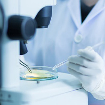
Microscopy methods start with inspection of the contaminated sample via optical microscopy using a stereo-microscope. If the contaminated sample appears the same as a clean control sample, then bulk analysis methods are best. The fact that discrete particulate contaminants are not present can still be substantial information for establishing provenance, the source of the contamination. But in instances where contamination is found to consist of solid phase contaminant particles, a series of sample preparation methods can further isolate the contaminants from the product matrix and characterize the particles.
This early stage of optical microscopy inspection yields the most valuable evidence about the origin of the contaminant particles—their size, shape, how they are incorporated into the product, their color and consistency. All of this provides clues about not only the composition of the particles, but how and when they might have been introduced. Capturing photomicrographs of the particles to document these qualities frequently aids later analytical steps and helps determine final results.
The next step involves manually isolating representative particles from the sample matrix under a stereo-microscope. This step is critical because the materials the particles are comprised of cease to be trace components of a bulk sample, and instead become the major components of the analyte. Concentrating the contaminant materials allows the use of methods that might otherwise be considered insufficiently sensitive and thus, in particulate analyses, the restrictive terms of major, minor or trace concentration lose their meaning. Depending on the characteristics of the particles—whether they are metallic, fibrous, granular or elastomeric—particles may be divided into portions and mounted for:
- polarized light microscopy
- scanning electron microscopy with energy dispersive X-ray microanalysis
- microscopic transmission mode Fourier transform infrared spectroscopy
- Raman microspectroscopy
- micro-X-ray diffraction analysis
- transmission electron microscopy
- additional analytical methods
Alternatively, the particles may be analyzed serially, moving the particles from one mounting form to another. Although not absolutely essential, a very clean environment such as an ISO Class 5 or higher cleanroom prevents introducing new contaminants. Generally, only a few methods are required to completely identify the particles.
Microscopy in Practice
As an example, ingredients or final products commonly have black particles. Are the particles thermally degraded or charred ingredients? Rubber? Plastic? Metal wear? Mold? When the particles are a millimeter and smaller the distinction becomes a challenge. The Food Safety Modernization Act has empowered FDA to ask food producers to understand the origin of contaminants in their products, and a multiple microscopy approach is beneficial in this regard. If the black particles in this example are determined to be metal, the alloy or alloys with which the particles share elemental consistencies are easy to establish. So, if particles are consistent with 304 stainless steel alloy—and most of the processing machinery is composed of 316 stainless steel alloy—then the source of wear particles can be more easily traced. Sometimes, processing equipment is manufactured with parts not meeting their expected specification and wear particles may be very unique to the part. Likewise, plastic and rubber parts have telltale compositions or fingerprint-like internal heterogeneity. Nondestructive microscopy methods document these revealing characteristics which can then be used to establish what, where and when a contaminant or adulterant was introduced into the food production process.
Adding microscopy methods to a contaminant investigation protocol prior to using bulk analytical methods provides leads about the provenance of the adulterants, which are otherwise lost from a protocol utilizing bulk analytical methods alone. Although some bulk analytical methods have intrinsically lower detection limits, the process of isolating contaminant particles with microscopy methods offsets the needed instrumental sensitivity while also capturing valuable morphology, size and consistency information. Collecting a more complete description of the contaminants can help reveal both their origin and a solution to the contamination.
Craig S. Schwandt, Ph.D., is the director of Industrial Services at McCrone Associates, Inc.
How Do Microscopy Methods Reveal the Source of Trace Contaminants?